You must be signed in to read the rest of this article.
Registration on CDEWorld is free. You may also login to CDEWorld with your DentalAegis.com account.
Case Report
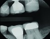
A 64-year-old woman presented with a chief complaint of food packing to the front of her implant. Clinical and radiographic examination revealed an existing Straumann Soft Tissue Level (RN) implant (Straumann, straumann.us) for tooth No. 19, restored with a prefabricated solid abutment and a PFM crown. The mesial marginal ridge of the PFM No. 19 was fractured, leaving an open proximal space between No. 19 and No. 20 (Figure 1).
Treatment options, risks, and benefits were discussed with the patient. Given the distalized position of the implant within the restorative space and the already developed soft-tissue contours generated by the existing restoration, a custom abutment was chosen to replace the existing solid stock abutment. A custom abutment allowed for not only better adaptation to the existing soft-tissue contours, but also much better support for the final restorative material, minimizing the risk for future ceramic fracture. In order to maximize how much of the edentulous space was filled with the abutment, a hybrid abutment was chosen. A monolithic IPS e.max crown (Ivoclar Vivadent, ivoclarvivadent.com) was chosen for the final restoration.
A Planmeca PlanScan scanner (Planmeca CAD/CAM Solutions, planmeca.com) was used to take a preoperative scan before removing the existing PFM No. 19, in order to fabricate a provisional restoration. Following removal of the PFM, the existing solid abutment was removed and replaced with an intraoral scan body (Straumann). Digital impressions were taken of the lower (Figure 2) and upper quadrant, after which the scan body was removed and a buccal bite scan was created with the teeth in occlusion. The previous solid abutment was then replaced and a provisional restoration was fabricated.
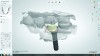
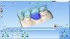
The case was sent digitally to Williams Laboratory in Gilroy, California, through DDX (Henry Schein, henryschein.com). Upon receiving the case, the laboratory imported it into CAD software (3Shape, 3shape.com) as well as Williams’ proprietary software. The zirconia hybrid abutment was designed via the Straumann DME (Figure 3) and the file split to simultaneously generate a printed 3D model with metal implant analog and to mill the zirconia portion of the hybrid abutment on a 5-axis mill. The 3D model was printed overnight while the zirconia abutment was sintered. The following day, the zirconia anatomic abutment was luted to its corresponding Straumann Variobase using Multilink (Ivoclar Vivadent). Before the case was shipped back, the abutment was placed on the 3D model and digitized, and the STL files were sent back to the dentist for in-office designing (Figure 4) and milling of the final e.max restoration with PlanCAD software and a PlanMill 40 (Planmeca CAD/CAM Solutions). The total time elapsed from when Williams received the case through DDX to when it was shipped back to the prescribing dentist was 24 hours.
Had this case utilized a custom titanium abutment, a 3D model with analog would have been unnecessary, and the design file for the abutment could have been sent back for restoration design and fabrication without any physical models or re-digitization required. Also, the restoration proposal split off from the titanium abutment could have been sent and milled in office, as Romexis software (Planmeca CAD/CAM Solutions) allows for open-architecture restoration STLs from any CAD software to be imported and milled on the PlanMill 40.
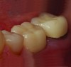
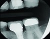
Thanks to the digitization of the final abutment, a monolithic e.max restoration was designed, milled, and ready for delivery prior to the office receiving the custom abutment. Upon delivery, the provisional restoration and solid abutment were removed. The new custom abutment was tried-in, along with the new e.max restoration to verify fit and occlusion. After the restoration was conditioned with Monobond Etch & Prime (Ivoclar Vivadent), the abutment was torqued to 35 Ncm, Teflon tape was placed in the screw access, and the restoration was cemented with SpeedCem (Ivoclar Vivadent) (Figure 5). Equigingival margin placement generated by the custom abutment design ensured thorough removal of excess cement. A post-operative radiograph was taken (Figure 6).
Conclusion
Digital dentistry is quickly making traditional clinician/laboratory workflows obsolete. For implant dentistry, it allows for significant savings in time and cost, while at the same time providing an optimal, patient-specific outcome. Furthermore, digital workflows give clinicians and laboratories much more flexibility, allowing each individual relationship to leverage the talent and resources that they have at their disposal to their maximum potential.
About the Authors
Bob Clark, CDT
Owner
Williams Dental Lab
Gilroy, CA
Clint Stevens, DDS
Private practice
Tulsa, OK