You must be signed in to read the rest of this article.
Registration on CDEWorld is free. You may also login to CDEWorld with your DentalAegis.com account.
Just as people are individuals and have unique features and personalities—including even identical twins—teeth are similarly unique. Each tooth is specific to a person. Teeth exist within a complex mastication system that consists of bones, soft tissues, muscles, ligaments, and nerves, and perform a vital function by allowing people to chew and eat. Teeth also have a significant esthetic component, especially evident when one smiles.
In recent years digital dentistry and computer-assisted design/computer-assisted manufacturing (CAD/CAM) have added new dimensions for the dental professional, including both dentists and laboratory technicians. However, the need to understand the fundamentals of anatomy, form, occlusion, and color science remains imperative. For dental professionals, understanding and mastering these fundamental concepts can be a lifelong learning process. The fact that each person and each tooth is unique makes this study a continual endeavor.
The US dental laboratory profession is experiencing a severe shortage of educated and trained dental technicians.1 Many laboratories are relying on CAD/CAM technology to offset this shortcoming. In some cases, the machinery is able to compensate for this deficiency. Many laboratories have segmented the process of creating a restoration into small steps in an effort to become more productive and profitable. In many cases the dental technician has become a step worker with little knowledge of the overall process. Laboratories are recruiting employees with technology interests who may not necessarily have any dental background. The technology has even enabled dental assistants to make restorations chairside, completely bypassing the dental laboratory. Technology has also made it possible to produce a restoration with little or no knowledge of why a tooth looks the way it does or why a tooth needs to be in a certain shape to function properly within the mastication system. These restorations appear toothlike but may be far from ideal. Thus, there is a great need to study and understand real teeth.
In this article, the size, shape, composition, and appearance of maxillary anterior teeth will be discussed from esthetic and functional perspectives. A total of 600 extracted maxillary incisors were studied: 200 each of central incisors, lateral incisors, and cuspids. The purpose of the article is to exhibit and discuss what makes teeth so unique and diverse.
Methods
During the process of this study, the author cleaned, photographed, and measured 600 teeth. Mesial distal crown width, labial lingual crown width, crown length, and overall tooth length were measured and recorded to the hundredths with digital calipers. Of the 600 teeth, 192 were photographed from four perspectives: facial, lingual, side, and occlusal. Labial and lingual photographs were taken in color. Labial/lingual photographs are displayed together to show how the lingual aspect can affect the shape of the incisal aspect. The labial/lingual images were made from separate images and combined using Photoshop. Side and occlusal views are displayed in black and white to put the focus on shape. All teeth were photographed with a dual-point source flash in a light box. The labial and lingual images were also photographed using cross-polarization to highlight internal color characteristics. Due to the vast number of photographs, only a select few are included in this article.
Anterior Tooth Size
Tooth measurements with histograms were taken (visit dentalaegis.com/go/cced953 to view the tooth measurements). A steep narrow histogram represented a similarity among tooth samples. For all tooth samples, there was a greater variety of overall length and crown length as compared to the mesial/distal and labial/lingual widths. Because the teeth were extracted, it was impossible to tell if this variety in length was due to incisal wear. The mesial/distal and labial/lingual measurements were nearly the same, making the teeth almost square in those dimensions. When viewing the tooth measurements (in the online graph), a blue vertical line represents the mean measurement, while a shaded area is one standard deviation. The measurements from these 600 maxillary incisors are similar to measurements reported by Woelfel2 taken in the 1970s.
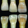
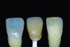
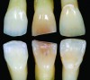
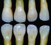
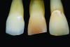
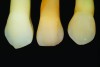
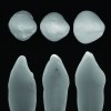
Figure 1 through Figure 3 show each tooth as represented in the histograms. The tooth in the middle of each set of these figures is the exact mean (and would be represented by the blue lines in the histograms). The left and right tooth in each set represent one exact standard deviation from the mean. From these images, the wide variety of tooth sizes and shapes that could be considered within normal measurements is evident, as these teeth are one standard deviation from the mean.
How Functional Component Affects Tooth Shape
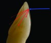
Anterior teeth are part of a complex chewing system, and the shapes of teeth play a role within this system. Often when people consider the shape of anterior teeth they refer to the incisal edge. More accurately, anterior teeth have an incisal ridge.3 This ridge consists of the labial incisal edge (Figure 4, red lines) and the lingual incisal edge (Figure 4, blue lines). These two edges form the boundaries of the incisal ridge. The lingual edge, which is often ignored, is the functional component that is dictated by the mandibular incisors and by how the patient chews. The labial edge is the esthetic incisal edge that is seen when a person speaks or smiles.
During chewing, the maxilla is stationary and the mandible moves. Described in Posselt’s Physiology of Occlusion and Rehabilitation in 1962,4 this envelope of mandibular movement is primarily dictated by the bones, muscles, and ligaments of the temporomandibular joint (TMJ). Dawson described how the teeth must work in an envelope of function within the mandibular movement.5 How the teeth occlude will influence their shape. The subject of countless theories, textbooks, seminars, academies, and study clubs, the science of occlusion is complex. Kois described this complex chewing system using three “P’s”: position, place, and pathway.6
The pathway, or how the teeth come together, is determined by the position of the mandible. The mandible’s position is determined by the bones, joints, muscles, and ligaments of the TMJ. The pathway the teeth take during chewing is partially defined by how the mandibular incisors function against the lingual surface of the maxillary incisors. The maxillary lingual incisal edge is the most critical part of the ridge (Figure 4) with regard to function. This lingual edge cannot be ignored or placed arbitrarily without consideration of the lower teeth. When the mandible closes during chewing, the lingual incisal edge of the maxillary teeth must not interfere with the pathway of the lower teeth. The labial incisal edge is the esthetic edge because it is what people see when a person smiles and talks. The location, or place, of the labial edge is mostly determined by facial esthetics and phonetics. The labial edge can be lengthened or moved as long as it does not affect function with the lower teeth, speech, and facial esthetics.
In an ideal situation the mandibular teeth work in harmony with the maxillary teeth. Different shapes and sizes of teeth cannot be interchanged without careful consideration of how the teeth will function within the chewing system. When restoring teeth, the lingual incisal edge must be considered for functional reasons prior to creating the labial incisal edge.
Optical Properties of Teeth
In order to understand the appearance of teeth, it is important to understand color, light, and how human beings see. Chu et al7 provided a thorough explanation of how light and color affect teeth and restorative materials. An understanding of these topics will enable dental professionals to better mimic teeth when making restorations and allow them to utilize a material’s strengths while recognizing and overcoming its weaknesses when choosing a restorative material.
Visible light is a form of electromagnetic energy.8 The electromagnetic energy spectrum ranges from cosmic waves to alternating current (A/C), and in the middle of the spectrum is a small portion known as visible light. The light that humans see is composed of different wavelengths, with the shortest visible light wavelength being violet (380 nm) and the longest being red light (700 nm). Wavelengths smaller than 380 nm and longer than 700 nm are not in the visible spectrum. Microwave and x-ray energy are just two examples of invisible electromagnetic energy.
When the wavelengths of light strike an object, they are viewed with the eye and recognized as a specific color in the brain. The type of light and composition of the object will dictate the color. This is why it is critical to understand color, light, tooth structure, and the composition of restorative materials.
White light has equal amounts of each wavelength within the visible spectrum.9 Light travels in a straight line and strikes an object at the angle of incidence. What happens to that light when it strikes the object depends on the type of light, the surface texture of the object, and the composition of the medium it is striking. All objects have different amounts of opacity, translucency, or transparency. The light will reflect back from completely opaque objects and pass through completely transparent objects. For translucent objects such as teeth, light will both reflect and pass through. This type of light is called refracted light. The way the light reacts within the medium is called scattering. To understand how light affects the appearance of teeth requires an understanding of reflected, refracted, and scattered light.
Reflected light—Reflected light enters the eye and hits the light-sensitive retina at the back of the eye. The retina contains rods and cones. The rods are responsible for vision at low light levels and do not determine color. The cones are responsible for a person’s color vision. Most people have 6-7 million cones that are concentrated on a 0.3-mm spot of the retina called the fovea centralis.
There are different types of reflected lights. A mirror reflection or specular reflection has the same angle of reflection as the angle of incidence (Figure 5). The surface texture of teeth is highly irregular (Figure 6) and will produce a diffuse reflection. A smooth, flat surface will produce a mirror reflection (Figure 5). Enamel surface texture varies greatly and affects the appearance of the teeth. Two restorations made from the same material may appear differently depending on the diffuse reflection. Surface texture on monolithic restorations is harder to control, which makes controlling the appearance more difficult.
Refracted light—As incident light traveling through space strikes a translucent object such as a tooth, it slows down and bends (Figure 7). Refracted light can be measured. The difference between the speed of light in a vacuum and the speed of light in a medium is called the index of refraction. This is explained by the equation n=c÷v, where “c” is the speed of light in a vacuum and “v” is the speed of light in a medium. The index of refraction for air is 1.0003; in contrast, the refractive index for a very dense material such as a diamond is a much higher 2.4.10
Scattered light—Light travels through a material in different ways, depending on the structure of the material. The light scattering within the material will depend on the material’s composition and the type of light. If the light is passing through a homogenous material, the light scattering will be consistent and predictable. Clear water, for example, has a very predictable and consistent structure. Its refractive index is 1.33. Not all translucent materials, however, are homogenous, or predictable. The refracted index alone is not the only factor. The scattering of light within a tooth will be highly irregular. Light will take an irregular path because of the composition of the tooth, the matrix (water), and the distance between these components. Some materials have more complex structures and matrices. Even air within the matrix will affect the scattering. The human tooth is a combination of liquid (water) and solids (enamel and dentin) as described below (in “Tooth Composition”). The solid portion of the tooth is a combination of several crystalline sizes and shapes in an aqueous matrix. The composition of teeth will scatter light in different ways because they are composed of a variety of different materials, each having its own refractive index.
Restorative dental materials have physical and optical properties that attempt to mimic the properties of teeth. Many modern monolithic ceramic materials have defined predictable crystalline structures that do not scatter light in the same way as a natural tooth. To compensate for this esthetic deficiency, a dental technician must layer different colors and opacities of material. Light scattering within homogenous monolithic materials makes the replication of teeth nearly impossible. In Figure 8 the natural tooth section on the right is 0.55 mm thick. From this cross-section it is easy to see the optical complexities of tooth structure. The feldspathic ceramic cross-section on the left is 1.5 mm thick (Figure 8). This cross-section shows the different layers of material that are necessary to mimic natural teeth. The sample in the center of Figure 8 is a replica of the left sample. It is made from monolithic zirconia. The zirconia cross-section shows the optical challenges a dental technician faces when using this material to match teeth. Monolithic materials have gained in popularity but present substantial esthetic challenges.
Tooth Composition
The composition of teeth can be seen in Figure 9. The coronal aspect of the tooth is composed of the pulp chamber, dentin, and enamel, while the root portion of the tooth consists of cementum, dentin, and the pulp canal. For the purpose of this article the cementum and pulp will not be discussed.
Enamel and dentin each has its own unique esthetic and mechanical properties. The outer portion of the tooth is covered by enamel, the hardest substance in the body.9,11,12 Enamel is highly mineralized, containing 96% to 98% inorganic substance. The most abundant mineral component (90%) is hydroxyapatite. Jenkins13 described enamel with 87% hydroxyapatite and 11% water. Kraus et al14 said enamel is made up of 90% hydroxyapatite and 4% water. Although the percentages differ, it is important to note that the enamel hard tissue is composed partially of water. The layer of the tooth directly beneath the enamel is dentin, which is softer and more flexible than enamel. It is also similar to, yet harder than, bone15 and is composed of 75% inorganic material and 20% organic material (mainly dentinal collagen). The remaining 5% is water and other materials.14 These are living tissues that are not static.
When people eat, their teeth move, flex, expand, and contract. Separately, these two materials—dentin and enamel—would not hold up to the stresses of the oral environment. Combined, their properties are quite extraordinary. The stiff outer enamel shell protects the softer, more flexible dentin core. This can be explained by the modulus of elasticity, which is the measure of an object’s ability to resist deformation when a force is applied to it. A stiffer material will have a higher elastic modulus. Enamel has an elastic modulus of roughly 80 GPa, while dentin has an elastic modulus of approximately 14 GPa.16,17 By comparison, zirconium dioxide (ZrO2), or zirconia, has an elastic modulus of 138 GPa; iron, 197 GPa; and diamond, 1035 GPa.18
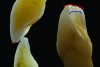
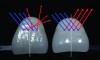
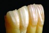
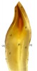
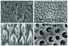
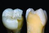
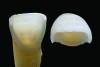
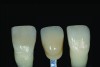
The critical mechanical properties of lithium disilicate, zirconia, enamel, and dentin also provide these structures with unique optical properties.17,19 Scanning electron microscopes (SEMs) of these materials (Figure 10) along with the tooth cross-section images (Figure 9) make it apparent that light will reflect, refract, and scatter differently for each material. According to ten Bosch and Coops,20 when incident light strikes a tooth it scatters sideward through the enamel from one to several millimeters. The enamel itself, as seen in Figure 11 through Figure 14, primarily acts to scatter light in the blue wavelength range. Very little color comes from the enamel, but the hydroxyapatite crystals perform a crucial esthetic function by scattering light. Scattering and absorption of light is much stronger within the dentin. The dentin is the predominant determinant of tooth color as seen in Figure 6 through Figure 9 and described by Zijp and ten Bosch.20
Figure 11 shows a cutaway of a posterior tooth, depicting the optical properties of dentin and enamel. Figure 12 is a central incisor with the dentin removed. Figure 13 shows two different teeth that have a similar color compared to an A2 shade guide. In Figure 14, the same two teeth as in Figure 13 are shown. Dentin has been removed from the tooth on the left, and enamel has been removed from the tooth on the right. It is evident from these images that the majority of the color comes from the inside layer of dentin.21 Very little color emanates from the enamel. The enamel acts primarily as a filter, or a scatterer, in terms of esthetics.20
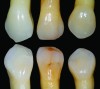
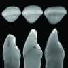
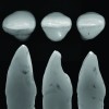
Shape and Appearance
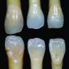
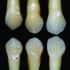
As previously explained, teeth shape and appearance are dictated by the anatomy of surrounding tissues, function, and tooth composition. The photographic element of this study provides an intimate appreciation of the diversity of teeth in terms of colors, shapes, and characteristics. The assorted images show the vast variety of shape and color. Figure 15 through Figure 17 show facial and lingual views, while Figure 18 through Figure 20 are captured using cross-polarization. Cross-polarization eliminates reflections and concentrates solely on color. Side and occlusal views are shown in black and white in Figure 21 through Figure 23 to highlight the shape of the teeth. These are the same teeth used in Figure 1 through Figure 3.
Discussion
Tooth shape and color is complicated, diverse, and fascinating. In an effort to elucidate it, and perhaps make learning easier, many textbooks have endeavored to highlight the commonalities of teeth.22 For example, some textbooks state central incisors are 10.5 mm long and 8.5 mm wide.2,23,24 Texts may show that central incisors have three mamelons and a white halo.23 Some textbooks use artists’ drawings that depict the author’s ideal shape of a tooth. With the increased popularity of CAD/CAM, it has become possible to choose a tooth shape from an artificial digital library. The technician simply picks a tooth from the library, makes it fit the empty space, and pushes a button. Then, either the restoration is colored with metallic oxides on the outside, or a small layer of veneering porcelain is added over the monolithic structure.
In reality, teeth have as many unique characteristics as they have commonalities. It is important to know and understand these commonalities, and they should be viewed as a good starting point—and not an ending point—when dentists and dental technicians restore teeth. With new materials being constantly developed, dental professionals must compensate by honing new skills to work with such materials.
Conclusion
Teeth play a vital role in people’s physical and emotional livelihood. As demonstrated in this study, in addition to the similarities among teeth, there is an immense diversity of teeth. Just like people, teeth have unique characteristics and individuality. Understanding this allows dental professionals to more effectively create esthetic restorations for patients.
Disclosure
The author had no disclosures to report.
About the Author
Steve McGowan, CDT
Owner
Arcus Laboratory
Kenmore, Washington
Queries to the author regarding this course may be submitted to authorqueries@aegiscomm.com.
References
1. Research indicates further decrease in laboratory numbers by 2017. Inside Dental Technology. 2015;6(8):6.
2. Fuller JL, Denehy GE, Schulein TM. Concise Dental Anatomy and Morphology. 4th ed. Iowa City, IA: University of Iowa College of Dentistry; 2001.
3. Nelson SJ, Ash MM. Wheeler’s Dental Anatomy, Physiology, and Occlusion. 9th ed. St. Louis, MO: Saunders Elsevier; 2010.
4. Posselt U. Physiology of Occlusion and Rehabilitation. Philadelphia, PA: F.A. Davis; 1962:52-55.
5. Dawson PE. Functional Occlusion: From TMJ to Smile Design. St. Petersburg, FL: Mosby; 2007:143.
6. Kois J, Hartrick N. Functional occlusion: science-driven management. Journal of Cosmetic Dentistry. 2007;23(3):54-57.
7. Chu SJ, Devigus A, Paravina RD, Mieleszko AJ. Fundamentals of Color: Shade Matching and Communication in Esthetic Dentistry. 2nd ed. Hanover Park, IL: Quintessence Publishing Co., Inc.; 2010:8-40.
8. Zwimpfer M. Color, Light, Sight, Sense: An Elementary Theory of Color in Pictures. Minneapolis, MN: Schiffer Publishing; 1985.
9. Zijp JR, ten Bosch JJ. Theoretical model for the scattering of light by dentin and comparison with measurements. Appl Opt. 1993;32(4):411-415.
10. Zajac A, Hecht E. Optics. 4th ed. San Francisco, CA: Pearson Higher Education, Addison-Wesley; 2003.
11. Chun K, Choi H, Lee J. Comparison of mechanical property and role between enamel and dentin in the human teeth. J Dent Biomech. 2014;5:1758736014520809. doi: 10.1177/1758736014520809.
12. Craig RG, Peyton FA. The micro-hardness of enamel and dentin. J Dent Res. 1958;37(4):661-668.
13. Jenkins GN. The Physiology and Biochemistry of the Mouth. Oxford, England: Blackwell Science Ltd.; 1978:54-112.
14. Kraus B, Jordan R, Abrams L. Dental Anatomy and Occlusion. Toronto, Canada, and Philadelphia, PA: B.C. Decker; 1988.
15. The glossary of prosthodontic terms. J Prosthet Dent. 2005;94(1):10-92.
16. Mange P, Belser U. Bonded Porcelain Restorations in the Anterior Dentition: A Biometric Approach. Chicago, IL: Quintessence Publishing Co., Inc.; 2002.
17. Bühler-Zemp P, Völkel T, Fischer K. Scientific Documentation IPS e.max® Press. Liechtenstein: Ivoclar Vivadent AG; 2011:7.
18. Richardson DW. Modern Ceramic Engineering: Properties, Processing, and Use in Design. 3rd Ed. Boca Raton, FL: Taylor & Francis; 2006:214.
19. Denry I, Kelly JR. Emerging ceramic-based materials for dentistry. J Dent Res. 2014;93(12):1235-1242.
20. ten Bosch JJ, Coops JC. Tooth color and reflectance as related to light scattering and enamel hardness. J Dent Res. 1995;74(1):374-380.
21. Hart PS, Hart TC. Disorders of human dentin. Cells Tissues Organs. 2007;186(1):70-77.
22. Linek HA. Tooth Carving Manual. Pasadena, CA: Wood and Jones; 1949.
23. Stavrianos C, Papadopoulos C, Vasiliadis L, et al. Enamel structure and forensic use. Research Journal of Biological Sciences. 2010;5(10):650-655.
24. Scheid RC, Weiss G. Woelfel’s Dental Anatomy. 8th ed. Philadelphia, PA: Wolters Kluwer/Lippincott Williams & Wilkens; 2012.