You must be signed in to read the rest of this article.
Registration on CDEWorld is free. You may also login to CDEWorld with your DentalAegis.com account.
The field of digital dentistry is undergoing a major transformation that is being driven by advancements that enhance precision, streamline workflows, and ultimately, improve patient outcomes. Among the most recent innovations in full-arch implant rehabilitation is intraoral photogrammetry, which is a cutting-edge technology that eliminates many of the limitations associated with traditional intraoral scanning methods.1 Although intraoral scanners have long been the standard for digital impressions, they can struggle with cumulative errors and distortions when capturing multiple implants in full-arch cases.2 The use of extraoral photogrammetry can improve accuracy in full-arch cases; however, these systems still present drawbacks and are often priced beyond the reach of many dental professionals.
For decades, clinicians have faced challenges in accurately capturing the spatial relationships of multiple implants. Traditional intraoral scanning relies on continuous image acquisition and stitching algorithms, which can introduce distortions when scanning across a large area, such as a full arch with multiple implants.3 Even small alignment errors between individual scans can lead to cumulative inaccuracies, and these inaccuracies can affect the fit of the final prosthesis and require extensive chairside adjustments. In addition, traditional intraoral scanning systems struggle to capture soft-tissue topography to maintain the needed fidelity for precise prosthetic intimacy. These limitations have made full-arch implant rehabilitation one of the most technically demanding procedures in dentistry.4 Intraoral photogrammetry overcomes these challenges by using coded scan markers that ensure precise implant positioning without relying on image stitching.
A Paradigm Shift in Total Record Accuracy
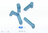
With intraoral photogrammetry technology, clinicians now have access to a highly accurate, efficient, and streamlined workflow for full-arch implant prosthetic planning. Unlike traditional intraoral scanning, intraoral photogrammetry employs specialized horizontal coded scan bodies that are placed over the implant multi-unit abutments to serve as precise reference points (Figure 1). Instead of relying on sequential image stitching, photogrammetry captures the exact 3D spatial relationships between the implants in a single step. This eliminates cumulative errors and provides a digital representation that is far more precise and reliable than that of conventional scanning techniques.
One of the most transformative aspects of intraoral photogrammetry is its ability to seamlessly transition from scan flags to scan bodies within a unified project file. Previously, clinicians needed to capture multiple separate scans at different stages of the treatment, which had the potential to lead to fragmented data and misalignment. With modern intraoral photogrammetry systems, the workflow is now all-in-one, allowing for easy conversion from high-accuracy horizontal coded scan bodies to users’ preferred scan bodies (Figure 2) without requiring additional scans. This feature simplifies the transition from surgery to fabrication of the prosthesis and ensures that relative implant and abutment positions remain consistent through the digital workflow.5
Enhanced Soft-Tissue Matching and Efficiency
Another major advantage of intraoral photogrammetry is its ability to accurately capture soft-tissue contours. Achieving an optimal emergence profile and soft-tissue adaptation is critical for full-arch restorations because the final prosthesis must integrate seamlessly with the surrounding gingival architecture. Traditional intraoral scanners often struggle to maintain accuracy when capturing soft tissue, especially in cases where the tissue dynamics change due to pressure from the scan bodies or movement during scanning. With intraoral photogrammetry, the precise implant positions are recorded without the need for direct soft-tissue alignment, which facilitates improved tissue-matching accuracy in the final restoration.
This soft-tissue precision plays a key role in the design and function of the prosthesis. By incorporating tissue-matching algorithms within the digital workflow, clinicians can ensure that the final restoration mimics the natural gingival contours, which reduces the need for excessive chairside adjustments and improves patient comfort. This is particularly important for all variations of fixed prostheses and implant-supported overdentures for which soft-tissue adaptation directly impacts esthetics and long-term stability.
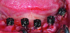
The introduction of coded healing caps has further improved the efficiency of this workflow. Traditionally, implant scanning has required the removal of healing abutments, the placement of scan bodies, and additional scanning steps before implant position data could be captured. With coded cap scan bodies, the implant positions are captured directly during the scanning phase, eliminating the need for additional scan body placement (Figure 3). The highly scannable shape, surface, and coding of coded healing caps reduces chair time, minimizes errors, and streamlines the transition from healing to design of the final prosthesis—even in a harsh, bloody soft-tissue environment.
Integrating Facial Scanning for Optimal Esthetics
Although intraoral photogrammetry can help to ensure precision in implant positioning, facial scanning can play an equally important role in achieving natural-looking full-arch restorations.6 Traditional workflows have relied on 2D facial photography and analog facebows to determine midlines, lip support, and smile dynamics. These conventional methods only offer a frontal view, without the ability to oscillate the head for maxillary occlusal cant analysis as well as anterior-posterior analysis. This limitation introduces variability and fails to present the full 3D relationship between the proposed prosthesis and the patient’s facial anatomy.7
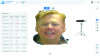
With modern 3D facial scanning, clinicians can digitally map key facial landmarks, including the smile line and midline, as well as assess lip mobility and facial symmetry (Figure 4). One of the greatest advantages of integrating facial scanning is its ability to enhance digital smile design. It allows for real-time visualization of the prosthetic design within the patient’s facial structure, ensuring that the restoration aligns naturally with the patient’s overall appearance.8
The All-in-One Digital Workflow
To improve digital precision, intraoral scanning, intraoral photogrammetry, and facial scanning can be combined into a comprehensive workflow. The process of merging these datasets involves aligning intraoral scans, intraoral photogrammetry data, and facial scans into a unified digital project file (Figure 5). By using common reference points, such as natural teeth or temporary markers, digital software can accurately register all of the datasets together. This enables simultaneous surgical and prosthetic planning, which ensures that the implant positions, bone structure, soft tissue, and esthetics are all considered in a single digital environment.
When intraoral photogrammetry data are merged with intraoral and facial scans, clinicians and technicians can both preview the final restoration in 3D simulation software. This allows for adjustments prior to fabrication, which not only improves patient communication and treatment acceptance, but also reduces the need for multiple trial prostheses, streamlining the entire workflow. Furthermore, being able to view patients in a 3D profile may offer guidance regarding future tooth and flange positions.
This all-in-one approach is also advantageous in improving multidisciplinary collaboration. Oral surgeons, prosthodontists, and dental technicians can all work from the same digital dataset, ensuring that both the surgical and prosthetic goals are aligned. This helps to reduce surgical complications, minimize prosthetic errors, and facilitate more predictable outcomes.
The following case report demonstrates how this integrated digital approach can transform clinical workflows to provide a more efficient and precise solution for complex full-arch restorations.
Conclusion
By eliminating the inaccuracies of traditional intraoral scanning, intraoral photogrammetry allows clinicians to achieve unprecedented levels of precision. When integrated with facial scanning, this technology ensures that final restorations are both functionally precise and esthetically natural looking, reducing the need for chairside modifications and enhancing patient satisfaction.
With intraoral photogrammetry’s advanced scan-matching capabilities, the transition from surgical placement to prosthetic design is seamless, accurate, and significantly faster than traditional workflows. The ability to capture implant positions, match scan flags to the surgical guide, and integrate soft-tissue data within a single project file—and when necessary, seamlessly transition from coded healing caps to final scan bodies—optimizes efficiency, which ultimately reduces chair time and improves the overall patient experience. As the technology continues to evolve, intraoral photogrammetry may become the gold standard in full-arch implant rehabilitation, offering a fully digital, highly predictable, affordable, and patient-friendly solution for modern implant dentistry.
Isaac Tawil, DDS, MS
Founder and Co-Director
Advanced Implant Education
Private Practice
Brooklyn, New York
Michael Erdos, DDS
Private Practice
Brooklyn, New York
References
1. Tawil I, Ganz SD, Pozzi A. Intraoral photogrammetry: the next step in full arch implant precision. International Magazine of Digital Dentistry. 2024;4:42.
2. Tawil I, Domingue D. Navigating the complexities of digital full-arch implant treatment: strategies to improve accuracy and deliver optimal outcomes. Inside Dentistry. 2024;20(6):33-40.
3. Tawil I, Ganz SD. Fully digital full arch? Continued advancements in full-arch implant restorations. Dentistry Today. 2023;42(4):50.
4. Mangano FG, Hauschild U, Veronesi G, et al. Trueness and precision of 5 intraoral scanners in the impressions of single and multiple implants: a comparative in vitro study. BMC Oral Health. 2019;19(1):101.
5. Jensen OT, Ross D, Jivraj S, Tawil I. Provisional prosthetic outcome when using photogrammetry for complete arch oral implants: a report of 111 patient treatments. Oral Maxillofac Surg Clin North Am. 2025;37(2):179-192.
6. Pozzi A, Arcuri L, Moy PK. The smiling scan technique: facially driven guided surgery and prosthetics. J Prosthodont Res. 2018;62(4):514-517.
7. Tawil I, Domingue D, Ganz SD. Digital full-arch maxillary rehabilitation. Inside Dentistry. 2024;20(7):28-35.
8. Mai H-N, Win TT, Tong MS, et al. Three-dimensional morphometric analysis of facial units in virtual smiling facial images with different smile expressions. J Adv Prosthodont. 2023;15(1):1-10.
9. Ganz SD, Tawil I. Full-arch implant surgical and restorative considerations: utilizing a full template guidance technique. Dentistry Today. 2019;38(9):72-78.